Structured-light surface scanning system to evaluate breast morphology in standing and supine positions

Measuring breast volume in hypertrophy: laser scanning or water displacement?

3-D breast area and breast area difference (BAD) calculation in cm 2 on

Early detection of the breast cancer using infrared technology – A comprehensive review - ScienceDirect
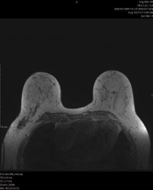
Magnetic Resonance Mammography: Practice Essentials, Technique, Preparation

Completeness of breast surface map for the three scanning positions.

Assessment of print quality: (a) Reconstructed CT model. M1, M2, M3

Applied Sciences, Free Full-Text
Tomography, Free Full-Text

Mamadou DIOP, Professor (Assistant), PhD Physics (Optics), The University of Western Ontario, London, UWO, Department of Medical Biophysics

Breast volumes of human subjects in three scanning positions.
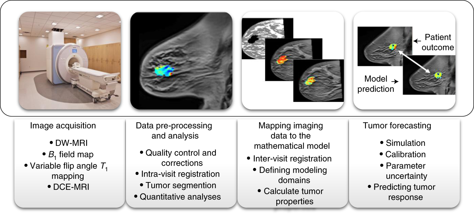
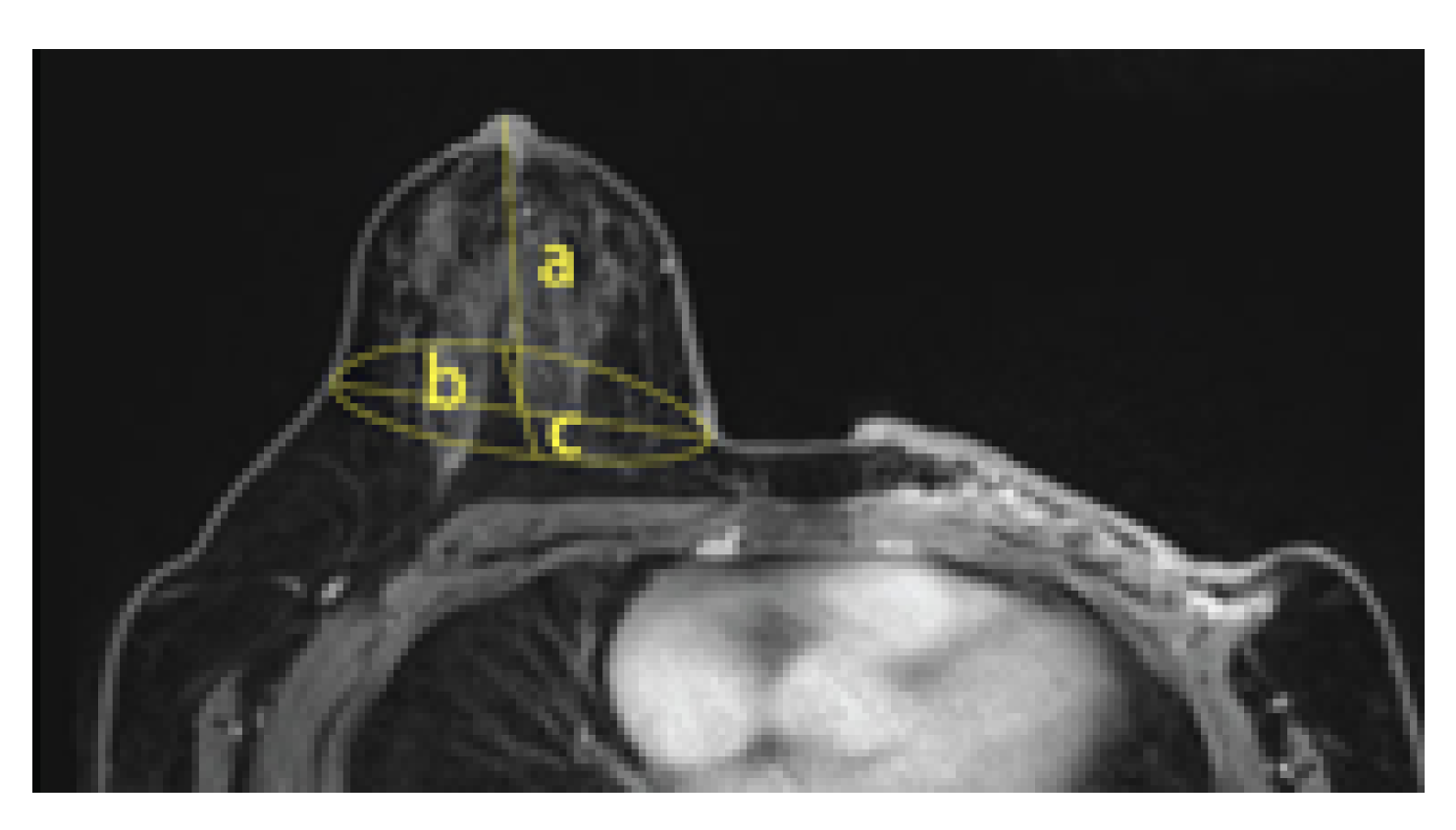
Quantitative magnetic resonance imaging and tumor forecasting of breast cancer patients in the community setting