Figure 3 from Descriptive anatomy of the interscalene triangle and
$ 10.99
4.7(68)In stock
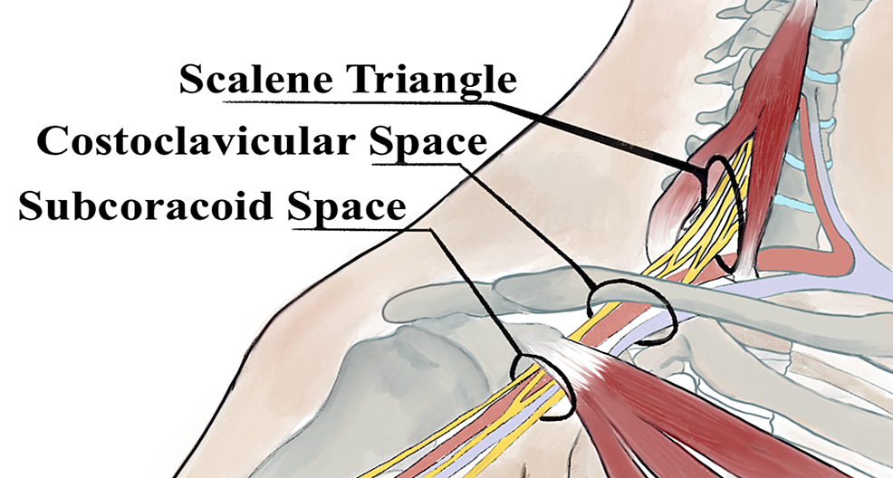
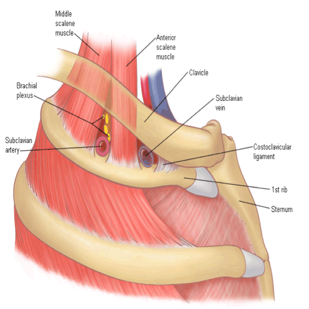
Fig 3. Depiction of the costoclavicular space. The neurovascular elements of the costoclavicular space can be seen here traveling superior to the first rib and inferior to the clavicle. The arrow indicates where measurements were taken. - "Descriptive anatomy of the interscalene triangle and the costoclavicular space and their relationship to thoracic outlet syndrome: a study of 60 cadavers."
Thoracic outlet syndrome: diagnostic and therapeutic update - ScienceDirect
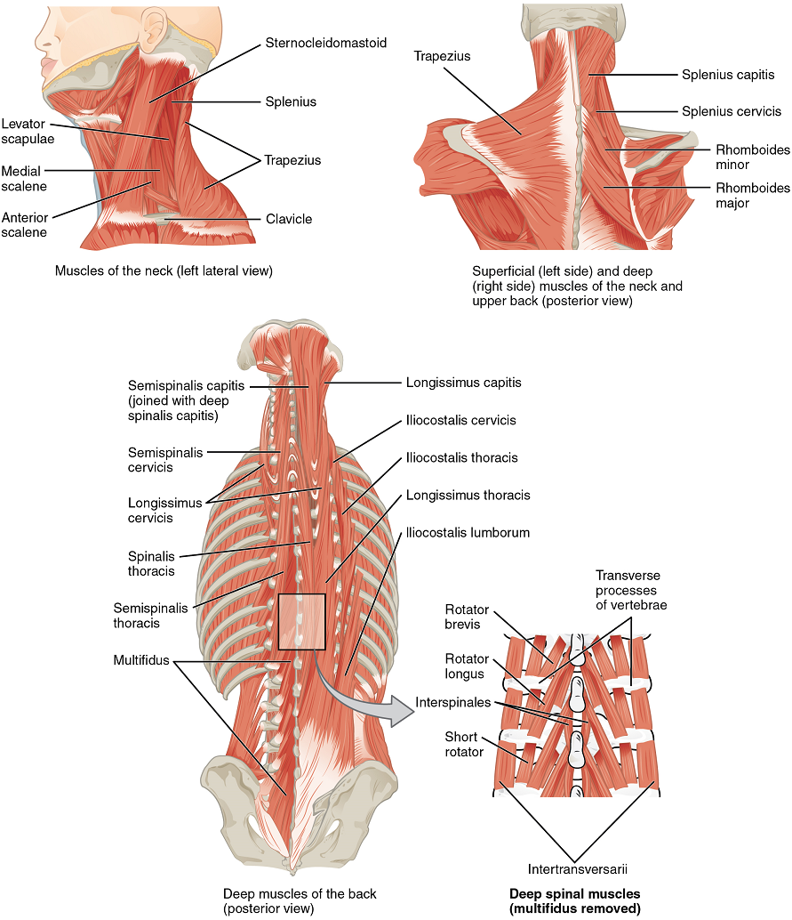
Chapter 13. Muscle Anatomy and Movement – Human Anatomy and Physiology I
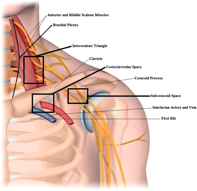
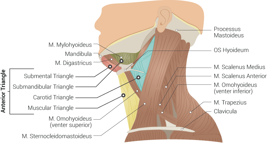
Anatomy, Head and Neck: Inter-scalene Triangle, Treatment & Management


/images/vimeo_thumbnails/297907323/5rW9QulJRmGVbpv5dLLgjw_overlay.jpg)

/images/vimeo_thumbnails/258305432/E2f2KldRHiRbDmuH6V6Cg_overlay.jpg)