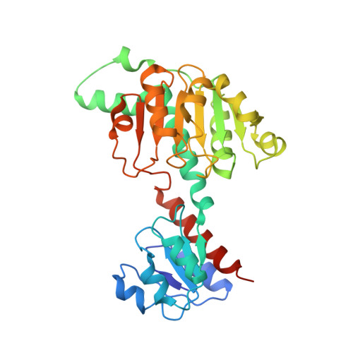
RCSB PDB - 2PRH: The structures of apo- and inhibitor bound human dihydroorotate dehydrogenase reveal conformational flexibility within the inhibitor binding site
Essentials-of-Paleomagnetism/EPSFiles/diags.eps at master · ltauxe/Essentials-of-Paleomagnetism · GitHub
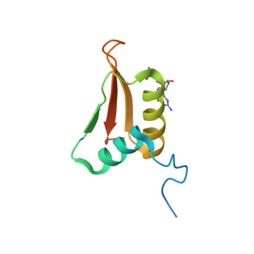
RCSB PDB - 2LRR: Solution structure of the R3H domain from human Smubp-2 in complex with 2'-deoxyguanosine-5'-monophosphate
RCSB PDB - 2PRH: The structures of apo- and inhibitor bound human dihydroorotate dehydrogenase reveal conformational flexibility within the inhibitor binding site
15.jpg
foto_2.jpg
Dihydrofolate Reductase human recombinant buffered aqueous glycerol solution Sigma
RCSB PDB - 2PRH: The structures of apo- and inhibitor bound human dihydroorotate dehydrogenase reveal conformational flexibility within the inhibitor binding site
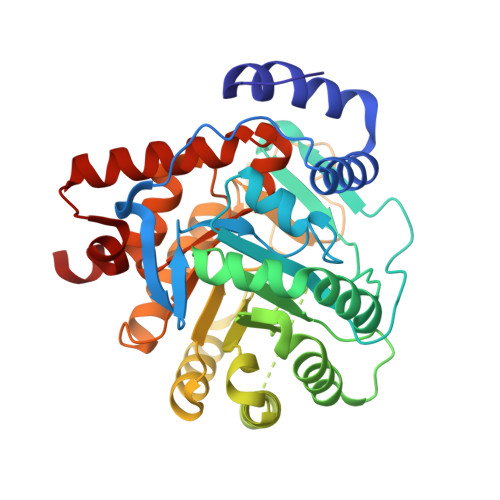
RCSB PDB - 6RJ2: Crystal structure of PHGDH in complex with compound 40
RCSB PDB - 6RJ2: Crystal structure of PHGDH in complex with compound 40
RCSB PDB - 2PRH: The structures of apo- and inhibitor bound human dihydroorotate dehydrogenase reveal conformational flexibility within the inhibitor binding site
DCP07932.JPG
RCSB PDB - 2PRH: The structures of apo- and inhibitor bound human dihydroorotate dehydrogenase reveal conformational flexibility within the inhibitor binding site