A) Preoperative intraoral periapical (IOPA) radiograph of 36. B) Post operative (IOPA) radiograph of 36. C) 1 month follow up IOPA radiograph of 36. D) 6 months follow up IOPA radiograph of
$ 24.50
4.7(291)In stock
A) Preoperative intraoral periapical (IOPA) radiograph of 36. B) Post operative (IOPA) radiograph of 36. C) 1 month follow up IOPA radiograph of 36. D) 6 months follow up IOPA radiograph of 36. E) 1 year follow up IOPA radiograph of 36. - IP Indian J Conserv Endod - clinical and preclinical conservative /restorative de
Type III apical transportation of root canal. - Abstract - Europe PMC
JaypeeDigital
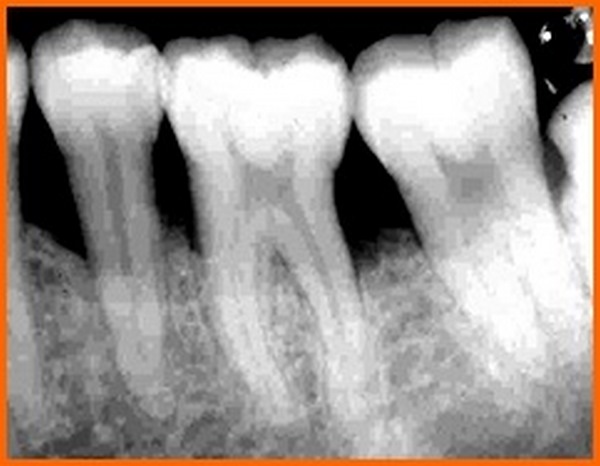
Postoperative radiograph revealed tooth 36 after root canal therapy
Pre-operative X-ray: suggested or obligatory - Style Italiano Endodontics
A) Preoperative intraoral periapical (IOPA) radiograph of 36. B) Post
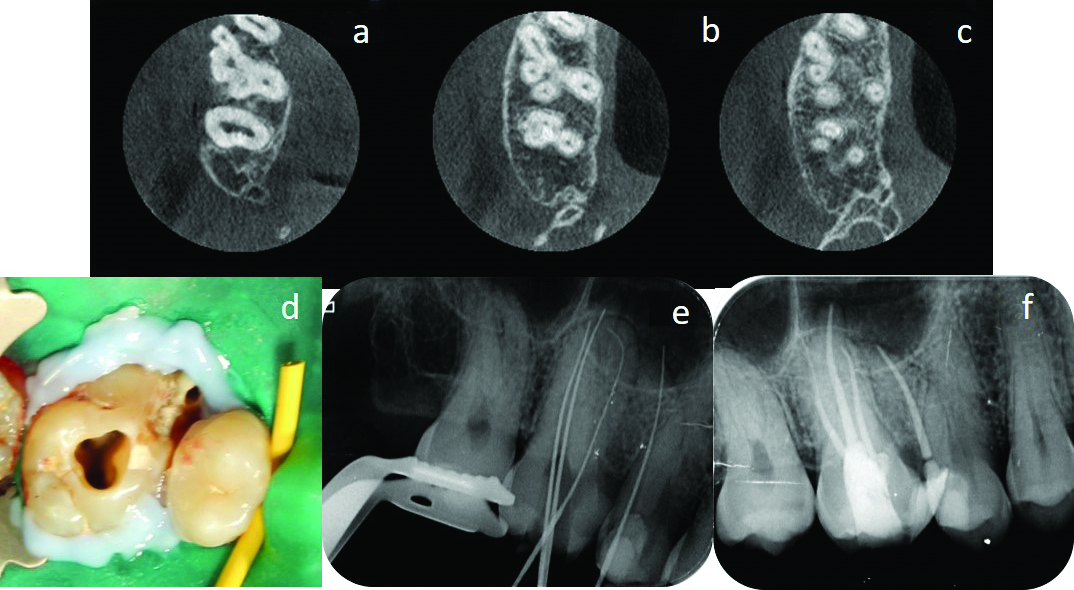
a) Preoperative IOPA radiograph of tooth #36. (b) Intraoral image
Preoperative and postoperative intraoral radiographs a: Preoperative
A Preoperative intraoral peri-apical (IOPA) radiograph of lower right
Coatings, Free Full-Text
File:Intraoral Periapical Radiograph (IOPA) showing Deciduous(Milky or Primary) Tooth 75 and developing crown of Permanent or Secondary Teeth 35, 36 and 37.jpg - Wikipedia
Effectiveness of Platelet Rich Plasma and Bone Graft in the Treatment of Intrabony Defects: A Clinico-radiographic Study